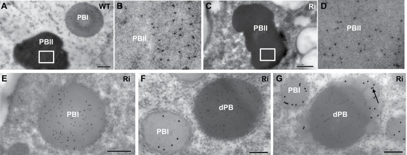
Fig. 9.
Immunocytochemistry of glutelin and prolamin in protein bodies. (A, B) Wild type; (C–G) OsSar1abc RNAi transformants. (B, D) Enlarged images of areas inside the boxes in (A, C). Glutelin and prolamin antibodies were labelled with 5nm and 15nm immunogold particles, respectively. The black arrow indicates prolamin accumulated in the blebbing structures of dPBs. Scale bars=500nm in (A, C, E) and 200nm in (F, G).

