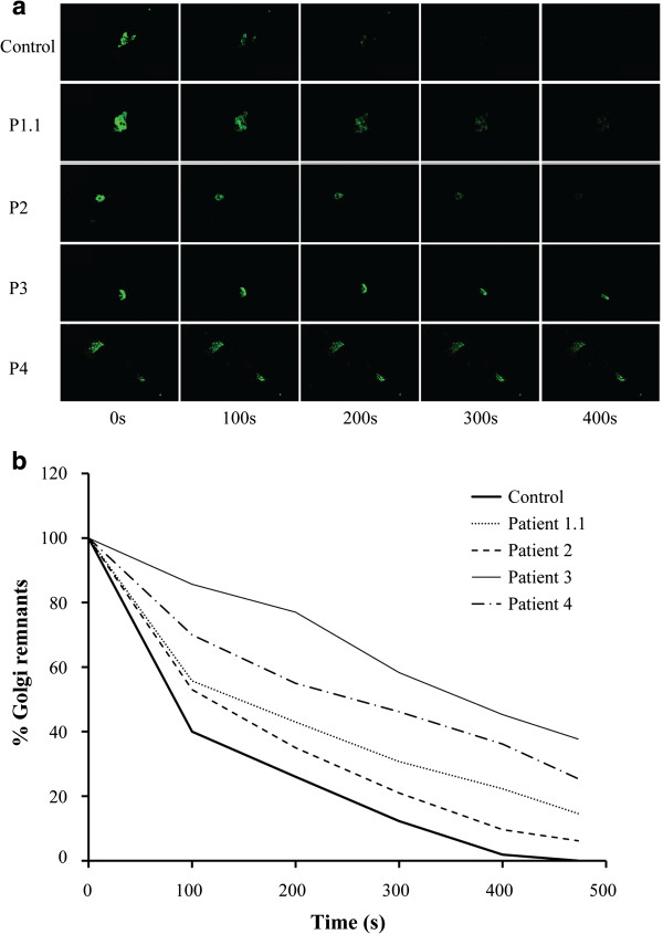
Figure 6.
BFA treatment of fibroblasts. Effects of BFA on control and COG5 deficient cells, visualized by time-lapsed video microscopy (a). Quantification of the remaining fluorescence (b). Note the delay in the redistribution of GalT-GFP from the Golgi into the ER in COG5-deficient cells compared to control.

