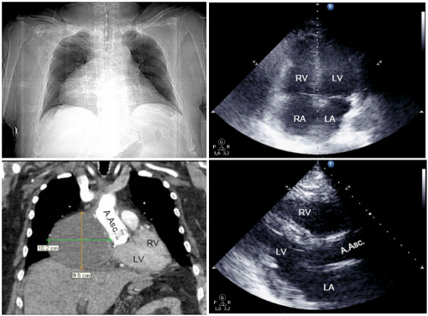
Figure 2.

Patient with cardiac tamponade on day 15 after ascending aortic replacement. Right atrial hematoma (102 × 95mm) not detected by transthoracic echocardiography (B-mode: apical 4-chamber-view right above, parasternal long axis right below). Thoracic CT-imaging with contrast medium, arterial phase (topography scan left above, coronary view, soft tissue window below left). Diagnostic imaging initiated after collapse necessitating cardiopulmonary resuscitation. LV left ventricle, RV right ventricle, LA left atrium, RA right atrium, A.Asc. ascending aorta.
