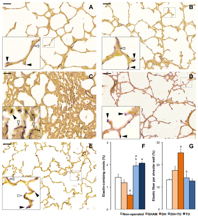
Figure 4. Pulmonary elastin pattern in the fetal rabbit model for CDH undergoing TO.
Miller’s elastic stain colored elastic fibers in black, and smooth muscle and cytoplasm in yellow. High-magnification insets show strong accumulation of elastic foci at the tips of secondary crests in the lungs of non-operated (A), SHAM (B), DH+ TO (D), and TO fetuses (E). DH lungs showed a disorganized pattern with predominant thick elastic fibers within the alveolar walls (C). Solid arrowheads indicate elastin-containing secondary crests. Open arrowheads indicate elastic fibers in the alveolar walls. F. The mean percentage of elastin-containing crests is decreased in DH lungs and restored by TO. G. The mean percentage of elastic fibers in the alveolar walls was increased in DH lungs and returned to SHAM values in DH+ TO lungs. Error bars represent the standard error of the mean, with 4 to 5 animals per study group. One-way ANOVA with Bonferroni correction was used for comparisons between surgical groups (SHAM, DH, DH+ TO, and TO) and unpaired Student’s t-test for comparisons between non-operated and SHAM fetuses. SHAM, sham-operated fetuses; DH, diaphragmatic hernia fetuses; DH+ TO, DH fetuses with tracheal occlusion; TO, sham DH fetuses with TO. a P < 0.05 vs SHAM; b P < 0.05 vs DH. Scale bars = 50 µm.

