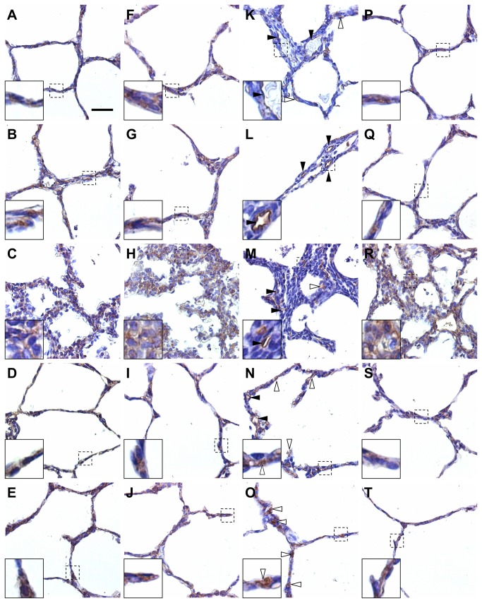
Figure 8. Localization of MMPs and TIMPs in alveolar septa in the fetal rabbit model for CDH undergoing TO.
Representative images of immunohistochemistry for MMP2 (A–E), MMP14 (F–J), TIMP1 (K–O), and TIMP2 (P–T) in non-operated (A, F, Q, P), SHAM (B, G, L, K), DH (C, H, M, R), DH+ TO (D, I, N, S), and TO lungs (E, J, O, T). MMP2, MMP14, and TIMP2 were expressed in alveolar epithelial cells in non-operated, SHAM, DH+ TO, and TO lungs (high-magnification inserts). MMP2, MMP14, and TIMP2 were also shown in mesenchymal cells in the thickened septa of DH lungs (diffuse staining in-high magnification inserts). TIMP1 was localized in endothelial cells of capillaries in all study groups (solid arrowheads), and in some alveolar epithelial cells (open arrowheads) in non-operated, SHAM, and DH lungs (K, L, M). DH+ TO (N) and TO lungs (O) showed a much more diffuse immune reaction for TIMP1 in alveolar epithelial cells (open arrowheads) as compared to non-operated and SHAM lungs. Scale bar = 50 µm in A through T.

