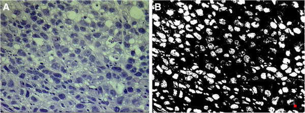
Figure 2.

Histologic section and binary image. (A) A hemotoxylin and eosin (H&E) stained image of the MCF-7 tumor and (B) the corresponding binary image indicating the presence of nuclei were identified using Image Pro Plus. The red square showed the area threshold used for counting the nuclei.
