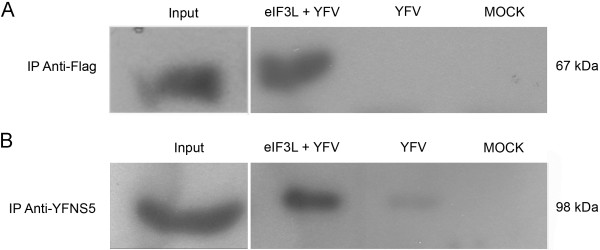
Figure 7.

Coimmunoprecipitation of YFV NS5 and eIF3L in mammalian cells. Vero cells were transfected with a plasmid expressing Flag-tagged eIF3L and infected with YFV strain 17D at an M.O.I. of 5. At 48 h post-infection, the cells were lysed and subjected to immunoprecipitation using anti-Flag M2 affinity gel beads. The coimmunoprecipitated proteins were analyzed by a western blot assay using an anti-YFNS5 antibody. (A) Cell lysates immunoprecipitated using the anti-Flag antibody. Input, positive control eIF3L (67 kDa) from transfected cells; eIF3L+YFV, eIF3L immunoprecipitated from transfected and infected cells; YFV, infected and non-transfected cells; MOCK, non-infected and non-transfected cells. (B) Coimmunoprecipitation of the NS5 protein with the anti-YFV NS5 antibody. Input, positive control YFV NS5 from infected cells (98 kDa); eIF3L+YFV, coimmunoprecipitation of YFV NS5 with Flag-eIF3L showing a positive interaction; YFV, MOCK, YFV NS5 was not precipitated in non-transfected cells.
