Fig. 1.
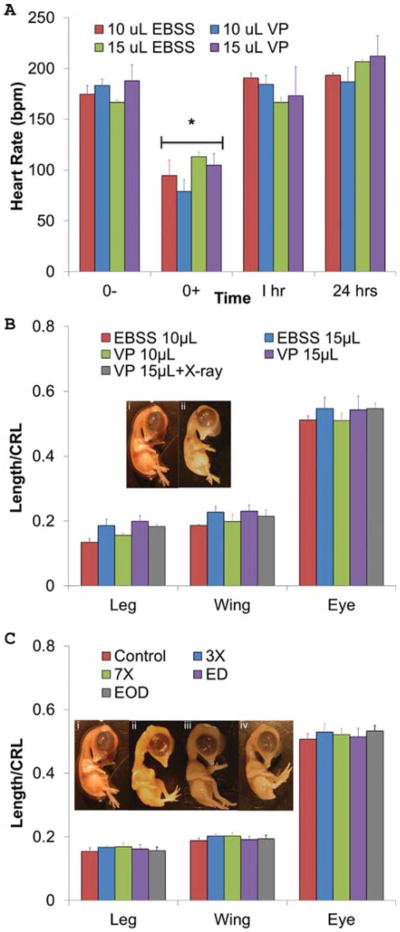
A: Acute embryonic heart rate adaptation to CT contrast microinjection. An acute drop in heart rate occurs after injection (0+) of VP and EBSS (P < 0.01), but heart rates return to initial levels within 1 hr. n = 3, 7, 3, and 6 embryos, L to R. B: VP causes no morphological defects. Day 10 limb length, wing length, and eye perimeter for each embryo were normalized by their respective crown-rump lengths (CRL). Legend indicates volume of either EBSS or VP injected. VP 15 μL + X-ray includes 1× X-ray (114 mGy of radiation). No significant differences were found. Day 10 embryos are (i) control and (ii) VP 15 μL + X-ray C: X-ray radiation causes no morphological defects up to 798 mGy doses. Identical measurements were taken as in Figure 1B. Treatments groups were as follows: imaged every day (ED), imaged every other day (EOD), imaged once on Day 4 at three times the dose (342 mGy, 3×), and imaged once at Day 4 at seven times the dose (798 mGy, 7×). There were no statistically significant differences in embryo morphology across limbs and X-ray treatments. Day 10 embryos are (i) control, (ii) ED, (iii) EOD, and (iv) 7×.

