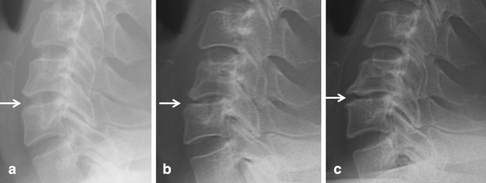
Fig. 3.

Lateral cervical radiographs obtained before (a) and at 1 (b) and 7 years (c) after C5/6 (arrows) AF in a 40-year-old male patient with soft disc herniation. This case showed remarkable postoperative reduction in disc height at the final FU. Aggravated disc degeneration is a routine radiological finding following AF. Early disc space narrowing (b) was followed by late spur formation (c). However, disc height reduction of the adjacent segments was not detected at 7 years and the segmental alignment had not changed markedly. This patient reported excellent results at the final FU
