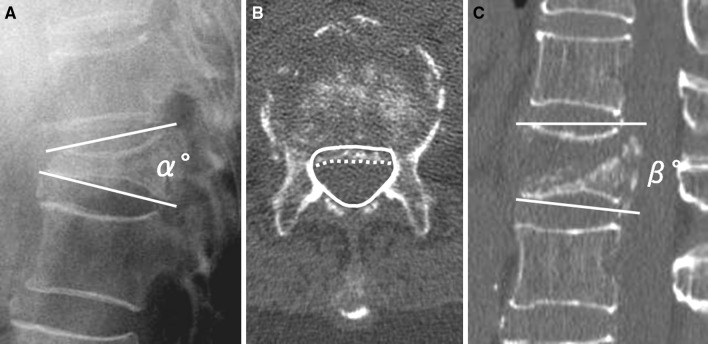
Fig. 2.
Three radiographic parameters demonstrating a “local kyphotic angle” (α°) measured using Cobb’s method with the patient in a sitting position and b “percent spinal canal compromise” calculated by dividing the area of intrusion by the total spinal canal area multiplied by 100. The total canal area is outlined by the solid line; the area of the retropulsed vertebral wall is demarcated by the dotted line. Areas of the spinal canal and retropulsed posterior wall are calculated from the total number of pixels per cross-sectional area (pixel/mm2). a, c “Intravertebral instability”, defined as the difference between local kyphotic angle on lateral radiography with the patient in a sitting position (α°) and that on a sagittal reformatted CT scan in a supine position (β°); α°–β°

