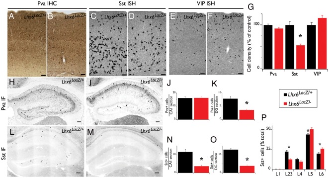
Figure 4.
Differentiation defects of cortical interneurons in Lhx6 hypomorphic animals. IHC of coronal brain sections from control (Lhx6LacZ/+; A) and Lhx6 hypomorphic (Lhx6LacZ/−; B) animals with antibodies specific for Pva. In situ hybridization of coronal brain sections from control (C and E) and Lhx6 hypomorphic (D and F) animals with a Sst (C and D) or a vasoactive intestinal peptide (VIP) riboprobe (E and F). (G) Quantification of the density of interneurons expressing the indicated markers from the somatosensory cortex of Lhx6 hypomorphic animals (red bars) relative to their control littermates (black bars); n = 3 animals per genotype. IF in the hippocampus of control (H and L) and of Lhx6 hypomorphic (I and M) animals with antibodies specific for Pva (H and I) or Sst (L and M). (J, K, N, O) Quantification of IF experiments; n = 5 animals per genotype. (P) An increased proportion of the Sst+ cells are located in the deeper layers of the neocortex of Lhx6 hypomorphs; n = 7 animals per genotype. All significant differences (P < 0.05) are indicated above the graphs with an asterisk. Scale bars 100 μm.

