Figure 5.

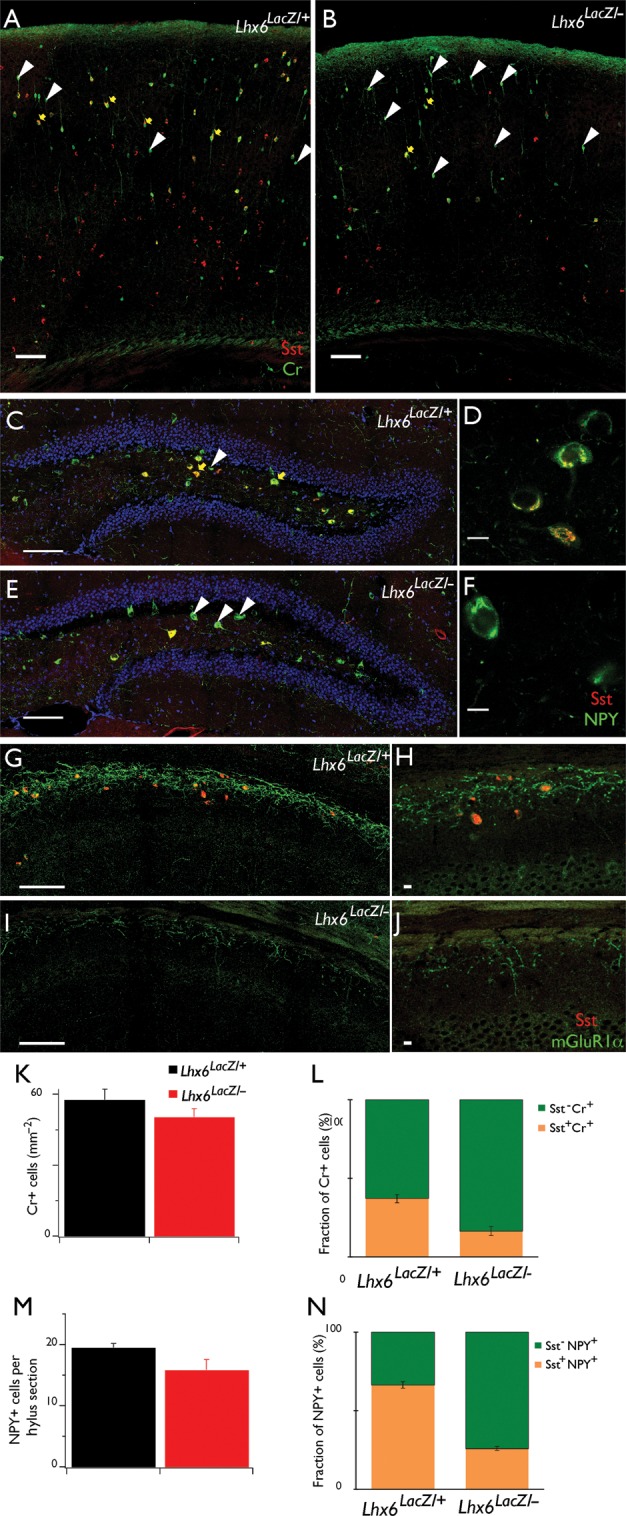
Partial differentiation of Sst+ interneurons in Lhx6 hypomorphic animals. Double IF of coronal brain sections from control (A) and Lhx6 hypomorphic (B) animals with antibodies specific for Cr (green) and Sst (red). No changes were observed in the total density of Cr+ cells (K), but the fraction of Cr+ cells that co-express Sst (yellow arrows in A and B, orange bars in L) is significantly reduced in Lhx6 hypomorphic animals, resulting in an increased fraction of Cr+Sst− cells (white arrowheads in A and B, green bars in L). Double IF of the DG hylus region from control (C) and Lhx6 hypomorphic (E) animals with antibodies specific for NPY (green) and Sst (red). No changes were observed in the total number of NPY+ cells (M), but the fraction NPY+Sst+ cells (yellow arrows in C and E, orange bars in N) is reduced in hypomorphs. (D and F) show single confocal sections of representative examples of NPY+Sst+ (D) and NPY+Sst− (F) cells at higher resolution. Double IF of the stratum oriens of the CA1 region of the hippocampus from control (G) and Lhx6 hypomorphic (I) animals with antibodies specific for mGluR1α (green) and Sst (red), showing reduced expression of mGluR1α in mutant animals. (H and J) show single confocal sections at higher resolution. Scale bars 100 μm (A, B, C, E, G, I) and 10 μm (D, F, H, J). In (C–F), cell nuclei are counterstained with 4',6-diamidino-2-phenylindole (DAPI) (blue), to reveal the granule cell layer.
