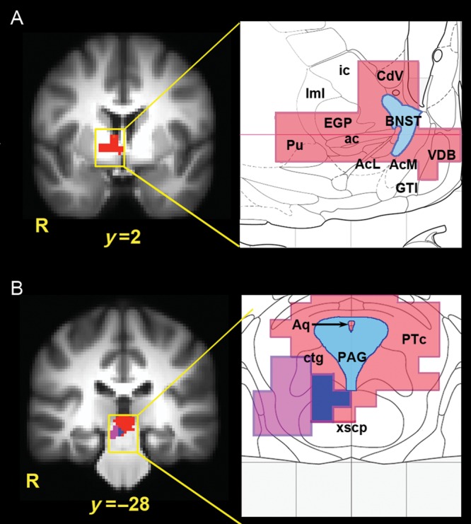
Figure 3.

(A) Activation of the right basal forebrain during sustained anticipation of aversion, as overlaid on an anatomical atlas (Mai et al. 1998). At P < 0.05 (corrected using Monte Carlo simulation), this activation included the dorsal and central BNST (shown in blue), as well as the external globus pallidus (EGP), ventral caudate (CdV), putamen (Pu), vertical limb of the diagonal band (VDB), great terminal island (GTI), and dorsal/posterior portions of the nucleus accumbens (AcL = lateral accumbens core; AcM = medial accumbens shell). ac, anterior commissure; ic, internal capsule, lml, external medullary lamina of the globus pallidus. (B) Sustained anticipation of aversion was accompanied by increased midbrain activity (red) overlapping with the anatomical location of the PAG (blue) and pretectal area (PTc) when overlaid on an anatomical atlas (Mai et al. 1998). PPI analysis revealed an overlapping midbrain cluster (purple) showing increased functional coupling with the anterior mid-cingulate cortex during sustained anticipation. All activations shown at P < 0.05 (corrected using Monte Carlo simulation). Aq, cerebral aqueduct; ctg, central tegmental tract; xscp, decussation of the cerebellar peduncle. Adapted from Mai et al. 1998.
