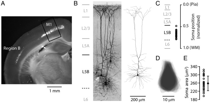
Figure 1.
Anatomical location and somatodendritic morphology of retrogradely labeled corticospinal neurons. (A) An epifluorescence image of a coronal brain slice prepared several days after injection of a fluorescent tracer into the cervical spinal cord, showing retrogradely labeled corticospinal neurons in layer 5B. Highest densities of labeled somata were found in 2 regions: Primary motor cortex (M1), and a separate lateral area (Region B). Box: Region in M1 where most recordings of M1 corticospinal neurons were made. (B) Dye-filled corticospinal neurons identified by retrograde labeling, whole-cell recorded with biocytin in the pipette, and imaged with 2-photon microscopy. Maximum intensity projection of image stacks. Laminar borders (indicated on the left) were measured and averaged from bright-field slice images (n = 60). The lower border of layer 5B was estimated from the lower edge of the distribution of bead-labeled corticospinal somata in fluorescence slice images (n = 30). Right: Reconstructed morphology of one of the corticospinal neurons (arrow). (C) Cortical depths of corticospinal neurons in the main data set plotted as the distance of the soma from the pia, normalized to the cortical thickness (pia to white matter, WM). (D) Soma tracings for the recorded corticospinal neurons. Tracings are centroid centered and aligned on the apical axis. (E) Cross-sectional areas of the somata (error bars: SD).

