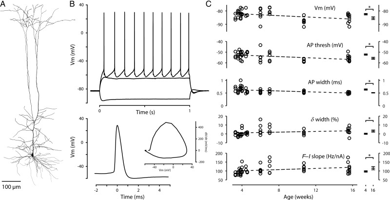
Figure 10.
Corticospinal intrinsic properties of older animals. (A) Reconstructed morphology of a corticospinal neuron from an older mouse (age P49). (B) Top: Responses to a family of injected steps of current (−300, +300, and +400 pA) recorded in the corticospinal neuron shown in (A). Bottom: Waveform of the first AP in the train shown at top. Inset: Phase-space representation of the same AP. (C) Intrinsic properties that varied with animal age. Each data point represents a single corticospinal neuron; the dashed line represents a linear fit to the data. The asterisk indicates a statistically significant (P < 0.05) difference between neurons from the original data set (∼4 weeks of age) and from the oldest animals (∼16 weeks of age). A few additional AP parameters differed between these age groups (see text). The δ width parameter is the change in AP width at approximately 10 Hz (percent difference from first to eighth AP).

