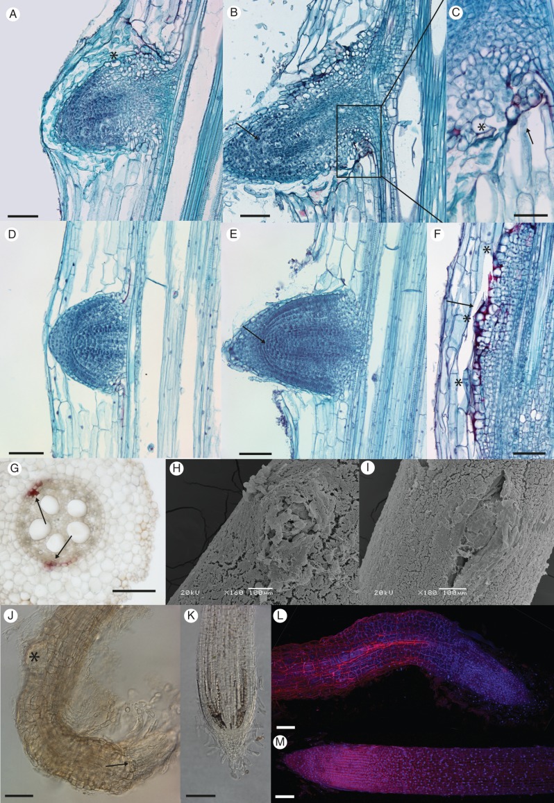
Fig. 3.
Changes in anatomy and emergence of lrt1 LRP. Plants were cultivated for 16 d in non-aerated hydroponics. LRP of the lrt1 mutant display an altered structure before (A) and after (B) emergence compared with the wild-type (D, E). Anomalously expanded highly vacuolated cells were observed on the surface and on the base of lrt1 LRP (A, C, asterisks). A loose arrangement of epidermal cells was obvious in the emerged lrt1 LRs (B). The structure of root apical meristem of LRP, and later in the apex of LRs with separation of root cap and tiers of initials and their derivatives is lost in lrt1 (B, arrow) compared with the wild-type (E, arrow). Lignifications (red) were often detected in lrt1 on the base of emerged LRP (C, arrow), around the malformed LRP (F, arrow; asterisks mark individual LRP), and in the pericycle of the primary root where initiation normally takes place (G, arrows). Emergence of LRs in lrt1 (H) and separation of the surface cell layers is modified; see (I) for the wild-type. The lrt1 mutant (J) shows irregular divisions in the surface area of LR tips and disorganization in the root apical meristem (J, arrow). Second-order LRP develop very close to first-order LR tips in lrt1 (J, asterisk), which often show strong curvature. Arrangement and anomalous cell volume growth is obvious in the epidermis of LRs (J, L, for lrt1; K, M, for the wild-type ). (A–F) Longitudinal permanent sections stained with Safranine and Fast Green. The sections were prepared from the first quarter of the length from the tip of the primary root. (G) Free-hand section from the middle of the primary root, HCl-phloroglucinol. (H, I) Scanning electron micrographs showing samples from the first quarter from the tip of the primary root. (J–M) Roots were cleared with NaI and stained with propidium iodide and Hoechst 33258. (J, K) Differential interference contarst microscopy; (L, M) confocal laser scanning microscopy, maximal projection. Scale bars = 100 µm, except (C) = 50 µm.

