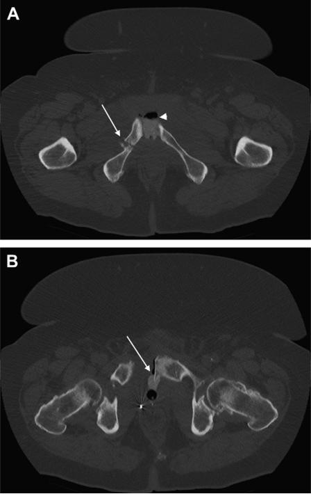
Figure 1.
Computed tomography scans of (A) pelvis with contrast showing pelvic fracture of right inferior pubic rami fracture (arrow), pubic diastasis, and extravasation of contrast and air (arrowhead) between pubic symphysis and (B) small bone exostosis extending posteriorly from left pubic symphysis into the bladder (arrow).

