Figure 1.
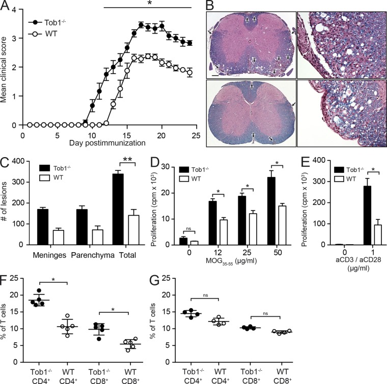
Tob1 deficiency exacerbates clinical and histological signs of EAE and increases myelin specific T cell responses. (A) Tob1−/− (n = 6) and WT (C57BL/6, n = 8) mice were immunized with MOG35-55 (these results are representative of three experiments). Mean disease score ± SEM is displayed. (B) Histology (Luxol fast blue–hematoxylin and eosin) of representative spinal cord samples. Meningeal and parenchymal inflammatory/demyelinating foci are indicated by arrows in Tob1−/− (top) and WT (bottom) mice. Bar: (left) 400 µm; (right) 80 µm. (C) Lesion quantification. (D) Proliferative responses in splenocytes of Tob1−/− and WT mice against MOG35-55. (E) Similar to D, but stimulation was performed with anti-CD3/anti-CD28. (F) Numbers of CD4+ and CD8+ spleen cells of MOG35-55 immunized (n = 4) Tob1−/− and WT mice. (G) Similar to F but with naive mice (n = 5). Experiments (C and G) are expressed as mean ± SD. *, P < 0.05; **, P < 0.01, Mann-Whitney U test. Data shown are representative two independent experiments.

