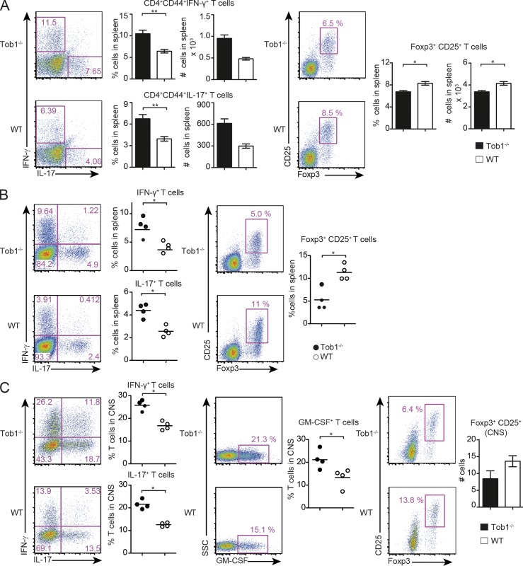
Figure 2.
Tob1 deficiency increases proinflammatory T cell responses. Tob1−/− and WT mice were immunized with MOG35-55. (A) Spleen cells were analyzed before disease onset for proportion and numbers of IFN-γ–, IL-17–secreting cells (gated on CD4+CD44+ cells, day 8), and for Foxp3 expression (CD25+Foxp+, day 7) by CD4+ cells. Representative FACS staining profiles are shown including quantification (Th1/Th17 n = 6; T reg cells n = 4 mice per group). (B and C) 14 d after immunization, spleen cells (B) and CNS infiltrating cells (C) were evaluated for secretion of IFN-γ, IL-17, GM-CSF, and for expression of CD25 and Foxp3 by CD4+ cells. The proportion of Foxp3+CD25+ cells (gated on CD4+) in the CNS of Tob1−/− and WT mice are shown in the rightmost panel. Representative FACS staining profiles are shown in each case including quantification (n = 4–5 mice per group). For all experiments, *, P < 0.05; **, P < 0.01, Mann-Whitney U test. Data shown are representative of two independent experiments. Error bars indicate SEM. Horizontal bars indicate mean.

