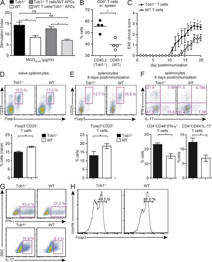
Figure 3.
Tob1 deficiency in T cells modulates T cell responses. (A) Whole spleen cells from immunized Tob1−/− and C57BL/6 mice were depleted of CD3+ cells and used as APC in co-culture with CD4+ T cells from Tob1−/− or WT T cells. 3H-thymidine incorporation in response to MOG35-55 is shown. (B) Rag1−/− mice were reconstituted with identical numbers of CD4+ cells from Tob1−/− (CD45.2) and transgenic WT (C57BL/6-CD45.1) mice. CD4+ T cells were isolated 12 d after immunization with MOG35-55 and quantified by FACS. Horizontal bars indicate mean. (C) Disease course after immunization with MOG35-55 of Rag1−/− mice reconstituted with CD4+ T cells from Tob1−/− or WT mice. Mean EAE disease score ± SEM is shown. (D and E) Proportion of splenic Foxp3-expressing cells (CD25+Foxp3+) by CD4+ cells in Rag1−/− mice either naive (D) or 8 d after immunization (E). (F) Proportion of splenic IFN-γ–, IL-17–secreting cells gated on CD4+CD44+ cells 8 d after immunization. (G) Polarization of T cells into a Th1 lineage was induced by IL-12 and polarization into a Th17 lineage was induced by IL-23, IL-6, and TGF-β. (H) Polarization of T cells into T reg cells was achieved with TGF-β. Intracellular cytokine staining for IFN-γ and IL-17 after 3 d in culture, and histograms for Foxp3 expression (gated on CD4+CD25+ cells) after 5 d in culture, are shown. Representative FACS staining profiles are shown including quantification (C, n = 4; D, n = 6; E, n = 4). For all experiments, data shown are representative of at least two independent experiments. *, P < 0.05, Mann-Whitney U test. Error bars indicate SEM.

