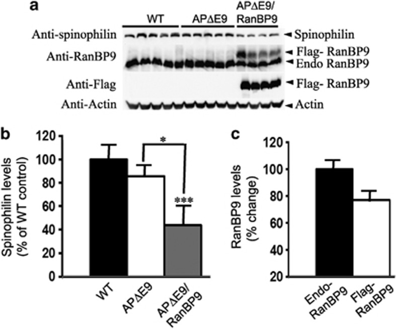
Figure 2.
RanBP9 overexpression in APΔE9 mice decreases spinophilin protein levels in the hippocampus at 12 months of age. (a) Brain homogenates from APΔE9 double-transgenic, APΔE9/RanBP9 triple-transgenic and age-matched WT control mice were subjected to SDS-PAGE electrophoresis and probed with anti-spinophilin antibody to detect spinophilin protein. Immunoblotting using RanBP9-specific monoclonal antibody detected both flag-tagged exogenous RanBP9 and endogenous RanBP9 in the triple-transgenic mice, while WT mice and APΔE9 mice showed expression of endogenous RanBP9 only (panel 2). Flag antibody detected expression of flag-tagged exogenous RanBP9 in APΔE9/RanBP9 triple transgenic mice only (panel 3). (b) Image J quantification showed decreased levels of spinophilin by 45% in APΔE9/RanBP9 mice compared with WT, but no change in APΔE9 mice. Compared between APΔE9 and APΔE9/RanBP9 mice, spinophilin levels further reduced by 34%. (c) Relative expression levels of endogenous and flag-tagged exogenous RanBP9 levels in the hippocampus of triple transgenic mice. Reduced levels of spinophilin were also confirmed by immunohistochemistry in the CA1 region of the hippocampus in the APΔE9/RanBP9 triple-transgenic mice compared with APΔE9 and WT mice. ANOVA followed by post hoc Tukey's test revealed significant differences. ***P<0.001 in APΔE9/RanBP9 mice compared with WT mice. *P<0.05 in APΔE9 versus APΔE9/RanBP9 mice. The data are mean±S.E.M., n=5 for WT and APΔE9 and n=4 for APΔE9/RanBP9 genotypes

