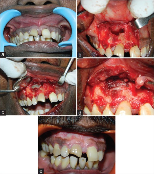
Figure 1.

(a) Pre-operative clinical picture (b) 15 mm × 16 mm × 16 mm gigantic lesion exposed along with root end resection being done (c) Combination of platelet rich fibrin (PRF) clot and hydroxyapatite crystals placed over the defect (d) 2 layers of PRF membrane placed covering the edge of the defect (e) post-operative clinical picture
