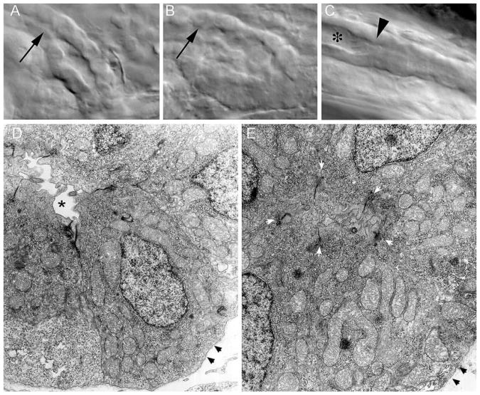
Figure 10.
Structural alterations in the distal pronephric nephron segment of pkd2 morphants. (A) Differential interference contrast images of the cloaca region of the pronephric duct in living, 3-dpf wild-type embryos show a patent lumen extending to the cloaca. (B) Polycystin-2 MOex3-injected embryos show an apparent collapse of the distal duct lumen. (C) Lumenal distension (*) immediately anterior to an area of duct occlusion (arrowhead in C) in a Polycystin-2 MOex3-injected embryo. Cilia beat and length appeared normal in areas of lumenal distension (data not shown). (H) Electron micrograph cross-section of the wild-type distal pronephric duct showing a patent duct lumen (*). For reference, cell basement membrane is denoted with arrowheads. (I) A similar region of the pronephric duct in a Polycystin-2 MOex3-injected embryos appears occluded by extended apical cytoplasm of duct epithelial cells. Five apical adherens junctions of the MOex3 duct epithelial cells can be seen (white arrowheads); however, the apical membranes of these cells abut with opposing cells, occluding the duct lumen. Black arrowheads, basement membrane.

