Figure 6.
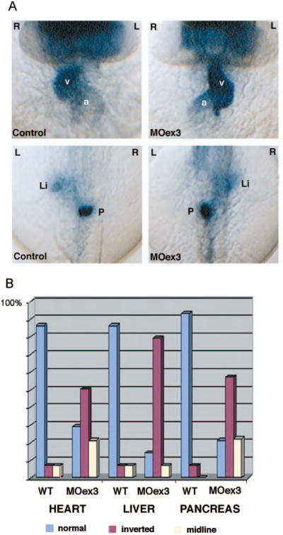
Polycystin-2 function in left–right asymmetry. (A) Expression of cardiac myosin light chain 2 (cmlc2) in control embryos (top left) demonstrates normal positions of the heart ventricle (v) and atrium (a) relative to the embryo midline. Polycystin-2 MOex3 caused inversion of left–right axes in 50% of injected embryos (top left). Forkhead2 (fkd2) and insulin gene expression revealed similar inversion of liver (li) and pancreas (p) situs, respectively (bottom). (B) Quantification of left–right asymmetry defects. Wild-type embryos (wt) show normal situs in most all cases with a low level of background laterality defects. Polycystin-2 MOex3-injected embryos show a high degree of left–right axis inversion (approximately 50%) as well as midline organ position for the heart, liver, and pancreas.

