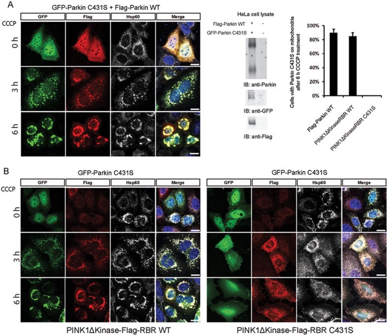
Figure 2.
Co-expression of WT Parkin restores Parkin C431S mitochondrial translocation.(A) Parkin C431S localizes to damaged mitochondria when co-expressed with WT Parkin. HeLa cells transfected with N-terminal GFP-Parkin C431S and Flag-Parkin WT, and then treated with CCCP. The cells were stained with anti-Flag antibody (red), anti-Hsp60 antibody (white) and Hoechst 33342 (blue). The relative levels of GFP- and Flag-tagged Parkin were determined by immunoblotting using anti-Parkin antibody; untransfected HeLa cells were used as control. The blot was also probed by anti-GFP and anti-Flag antibody, respectively. (B) A chimeric fusion of Pink1ΔKinase and Parkin RBR domain was sufficient to restore Parkin C431S mitochondrial localization. HeLa cells were transfected with GFP-Parkin C431S and Pink1ΔKinase-Flag-RBR WT or Pink1ΔKinase-Flag-RBR C431S, and then treated with CCCP and stained with anti-Flag antibody (red), anti-Hsp60 antibody (white) and Hoechst 33342 (blue). Scale bars, 10 μm. The graph shows means ± SD of percent of cells showing mitchondrial recruitment in three independent experiments, with 100 anti-Flag staining- and GFP-positive cells being counted per sample.

