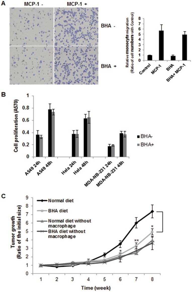
Figure 8.
BHA has no effect on MCP-1-directed monocyte migration and tumor cell growth. (A) Elutriated human monocytes were treated with MCP-1 (50 ng/ml) and the cell migration was assayed by using Transwell inserts. Cells were pretreated with or without BHA (100 μM) for 1 h prior to loading onto the migration chamber. Migrated cells were stained with crystal violet and evaluated by blind counting of the migrated cells on the lower surface of the membrane of 5 fields per chamber. Representative image is shown in the left panel and quantitative analysis is shown in the right panel. Data in monocyte migration detections represent the means ± SD of nine determinants from 3 independently prepared human samples each with 3 measurements. (B) A549, Hela and MDA-NB-231 cells were treated with or without BHA and the cell proliferation were detected by the MTT assay at 24 h and 48 h. The figure shows means ± SD from three independent experiments. (C) Tumor growth curve of MDA-MB-231-tdTomato cells inoculated into mouse mammary fat pads is shown. Female athymic nu/nu mice with or without macrophage depletion were divided into normal NIH-31 chow or NIH-31 chow with 7.5 g/kg BHA 1 week before the inoculation of tumor cells. Tumor size was measured for each animal every week starting from the day of the initial treatment. Relative tumor growth was normalized to week 1. Results are given as means ± SEM (n = 6/group). *P < 0.05; **P < 0.01 compared with normal diet control group at matched time point.

