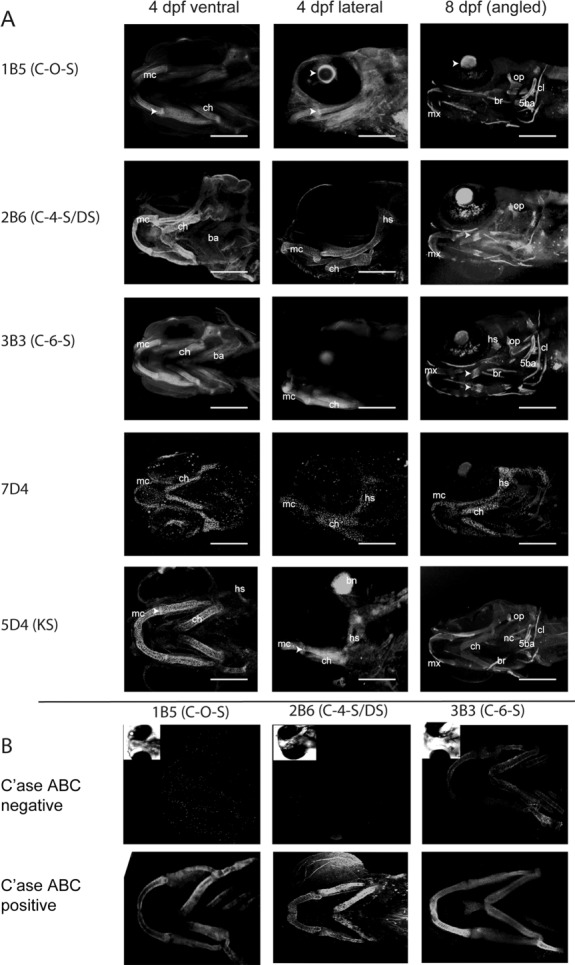
Fig. 3.

Chondroitin and keratan sulfate expression in the developing jaw. Confocal reconstructions showing comparative regions of the developing jaw of 8-day zebrafish larvae labelled with antibodies towards distinct chondroitin and keratan sulfate epitopes (refer to Table 2 for antibody specificities). In all images larvae are presented with anterior facing to the left. Arrowheads point to ossifying regions of the ceratohyal. ch, ceratohyal; mx, maxilla; mc, Meckel's cartilage; pq, palatoquadrate.
