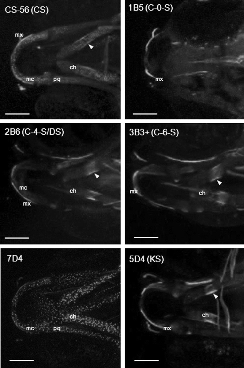
Fig. 4.

Chondroitin and keratan sulfate expression in the trunk and developing vertebral column. Immunohistochemistry of distinct chondroitin and keratan sulfate moities (as labelled) at 4 dpf and 8 dpf. All are lateral views with anterior to left. Trunk images at 4 dpf show a 4-somite span at the level of the cloaca. At 8 dpf, the images show an 8–9-somite span from the anterior end of the fish. Scale bars = 100 μm in all panels. hm, horizontal myoseptum; sb, somite boundary; sk, skin; nc, notochord; ha, haemal arch; na, neural arch; vb, vertebral body.
