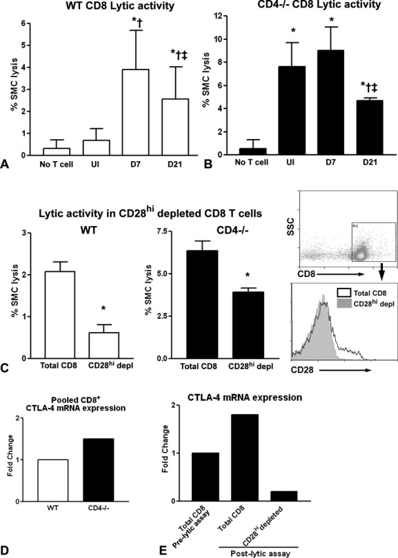
Figure 3.

CD8+ T‐cell cytolytic activity against syngeneic SMCs. CD8+ T cells were negatively isolated from spleens of uninjured (UI) WT (A) or CD4−/− (B) mice or 7 (D7) and 21 (D21) days after injury and cultured with target SMCs in a T‐cell to target ratio of 3:1. A, WT, n=8 to 17; *P<0.001 vs no T cells; †P<0.001 vs UI; ‡P<0.01 vs D7. B, CD4−/−, n=4 to 7; *P<0.001 vs no T cells; †P<0.01 vs UI; ‡P<0.01 vs D7. Cytolytic activity of CD8+ T cells after depletion of CD28hi from WT mice 7 days after injury (C, left) and uninjured CD4−/− mice (C, middle). Right panel shows histogram of CD8b+‐gated cells after CD28hi depletion. CD8b antibody is PE conjugated, and CD28 antibody is APC conjugated. WT, n=3 each; *P=0.001; CD4−/−, n=4 each; *P=0.002. CTLA‐4 mRNA expression was assessed in isolated CD8+ T cells pooled from WT or CD4−/− mice before lytic assays (D) and in CD8+ T cells pooled after lytic assays in whole CD8+ or CD28hi‐depleted CD8+ cells from CD4−/− mice (E). SMC indicates smooth muscle cell; WT, wild type; CTLA‐4, cytotoxic T‐lymphocyte antigen‐4; APC, allophycocyanin.
