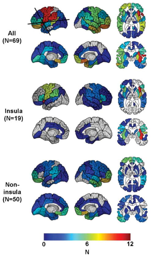
Fig. 1.
Number (N) of patients with lesion in each of the regions identified in this study, mapped onto a reference brain. Boundaries of anatomically defined regions are drawn on the brain surface. Regions names are provided in the Materials and Methods. Regions not assigned a color contained no lesions. (Top) All patients. The horizontal line marks the transverse section of the brain shown in the top row. The vertical line marks the coronal section shown in the bottom row. (Middle) Patients with lesions that involved the insula. (Bottom) Patients with lesions that did not involve the insula.

