
Figure 7. Canine arterial sections with thrombus exposed to fibrin-targeted or irrelevantly targeted nanoparticles.
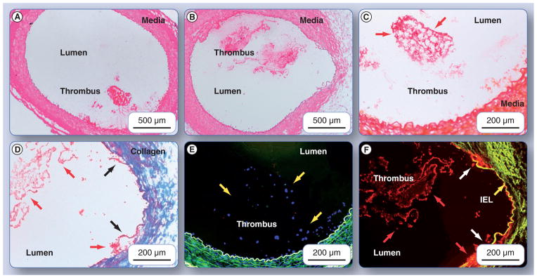
Hematoxylin and eosin-stained femoral artery segment from (A) a dog administered irrelevant-targeted urokinase-loaded particles, and (B) a dog given fibrin-targeted, urokinase-loaded particles, show anodal injury-induced thrombus attached to the femoral wall. Slice shown in (B) was fortuitously obtained near the needle injury site and revealed disruption of the intima, media and internal elastic lamina (highlighted with dark arrows [D & F]). Carstair’s staining of fibrin is shown in ([C & D]; which are magnified portions of [A & B], respectively), revealed an abundance of the target protein in both thrombi. Fibrin is shown in bright red, platelets in gray–blue to navy, collagen in bright blue, muscle in red and red blood cells in yellow. Note in (D) that the medial layer has been completely disrupted by the needle injury and collagen is primarily visible in this portion of the vessel wall. Fluorescent microscopic examinations of the thrombi that show the fibrin-targeted urokinase nanoparticles labeled with rhodamine (F) were bound along the serpentine surface of thrombus in a pattern consistent with the fibrin deposits revealed by the Carstair’s stain in (D). In contradistinction, only negligible binding of the irrelevant-targeted urokinase particles labeled with rhodamine was observed (E), despite a prominent accumulation of the target protein show in (C). IEL: Internal elastic lamina.