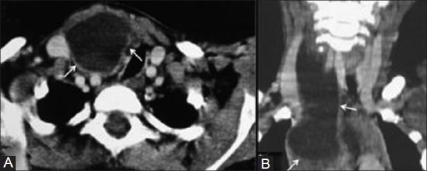
Figure 11(A, B).

Thymic cyst. (A) The axial contrast-enhanced CT scan shows a cystic lesion in the right side of the neck caudal to the thyroid gland displacing the trachea to the left. (B) The coronal reformatted image shows the lesion is parallel to the sternocleidomastoid muscle, extending into the upper mediastinum
