Figure 12.

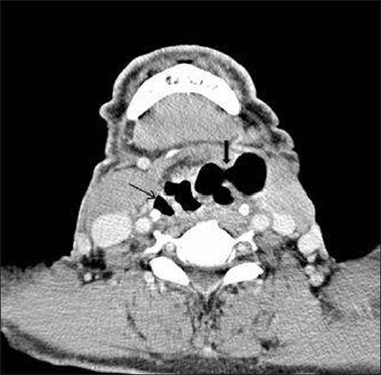
Laryngocele: Axial contrast-enhanced CT image shows air filled internal laryngocele (arrow) on the right and mixed laryngocele (arrowhead) on the left side

Laryngocele: Axial contrast-enhanced CT image shows air filled internal laryngocele (arrow) on the right and mixed laryngocele (arrowhead) on the left side