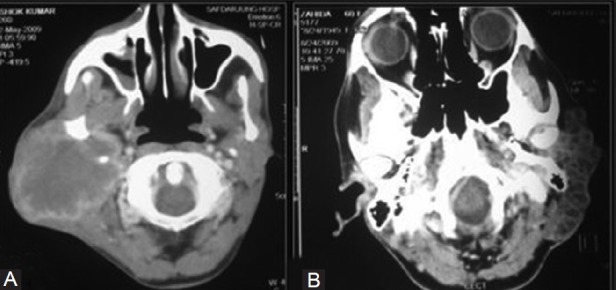
Figure 21(A, B).

Parotid masses. The (A) Axial contrast-enhanced CT scan shows a heterogeneously enhancing lesion in the right the parotid gland in a case of mucoepidemoid carcinoma. The (B) Axial CT scan in an adenoidocystic carcinoma showsa multi-cystic infiltrating lesion in the left parotid region
