Figure 9(A, B).

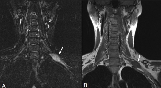
Coronal STIR and T1-weighted images demonstrate a well-defined fusiform lesion involving left brachial plexus. It is isointense on T1-weighted images and hyperintense on STIR images suggestive of nerve sheath tumor

Coronal STIR and T1-weighted images demonstrate a well-defined fusiform lesion involving left brachial plexus. It is isointense on T1-weighted images and hyperintense on STIR images suggestive of nerve sheath tumor