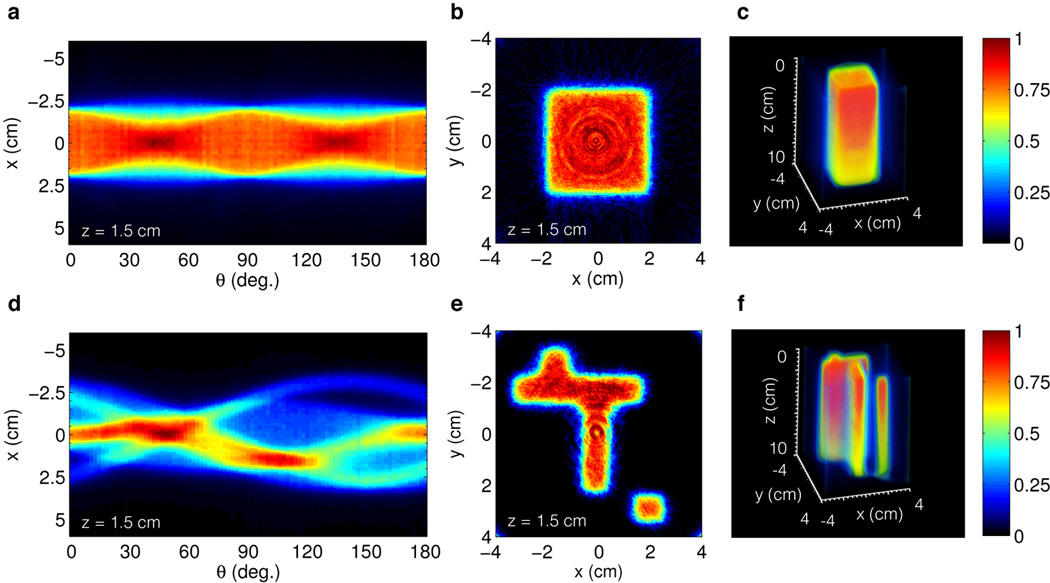
Fig. 2.

In (a,b) and (d,e) the sinogram and reconstructed cross section for Field A and B respectively. In (c) and (f) the full 3D reconstruction of Field A and B to a depth of z = 10 cm. The intensity decreases with depth due to exponential attenuation of the primary x-ray photon beam.
