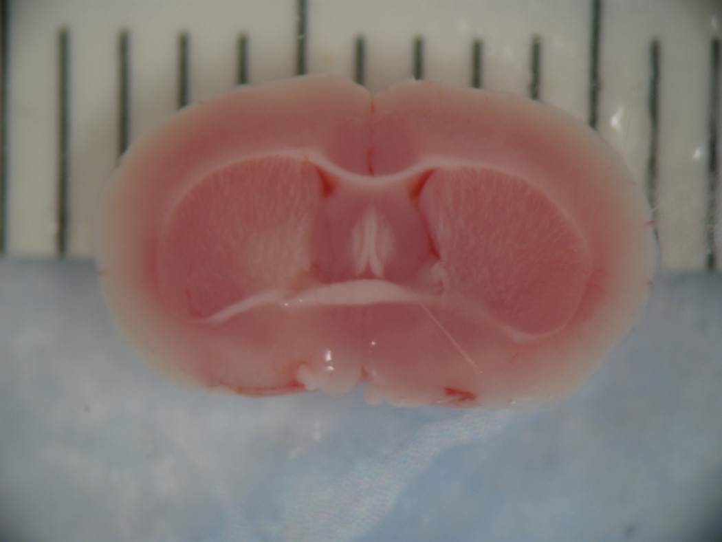
Figure 6.


On the left is a coronal section showing the caudal cerebral surface of the brain of a stroke animal. Nonstained tissue in the left hemisphere represents cerebral infarction in this coronal section stained with 2% triphenyltetrazolium chloride. On the right is a coronal section of the brain of an animal that underwent sham procedure. Note the uniformity in staining demonstrating a lack of cerebral infarction.
