
Figure 2.
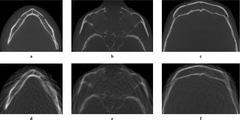
Examples of axial images, generated using a Scanora 3D (Soredex Oy, Tuusula, Finland). First row (a,b,c) shows reference images without motion artefacts. Second row (d,e,f) shows images with tremor artefacts. (a,d) Mental foramen; (b,e) infraorbital foramen; (c,f) supraorbital foramen