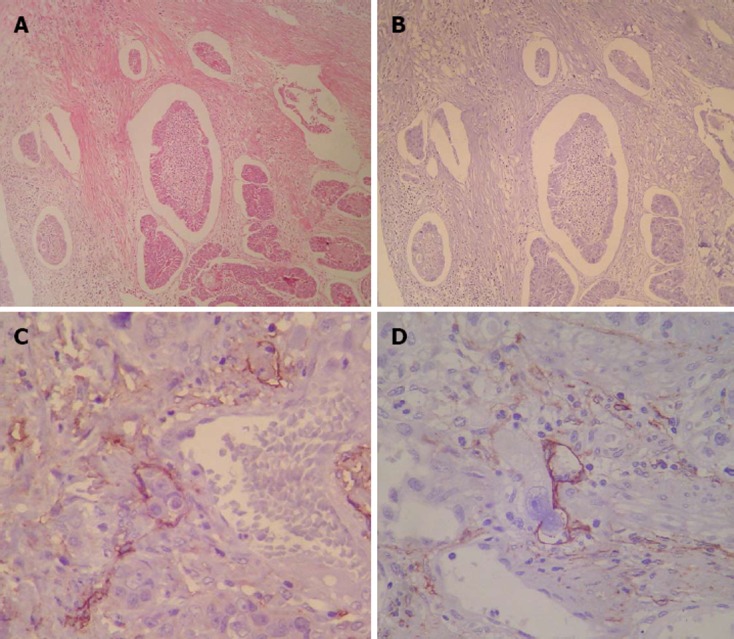
Figure 2.

Example of a patient diagnosed for lymphatic vessel invasion by routine histological examination. A: Example of a patient diagnosed as positive for lymphatic vessel invasion (LVI) by routine histological examination; B: As false-positive for lymphatic vessel invasion by D2-40 (× 100); C, D: Examples of patients diagnosed as free of LVI by routine histological examination. False-negatives for LVI detected by D2-40 (× 400).
