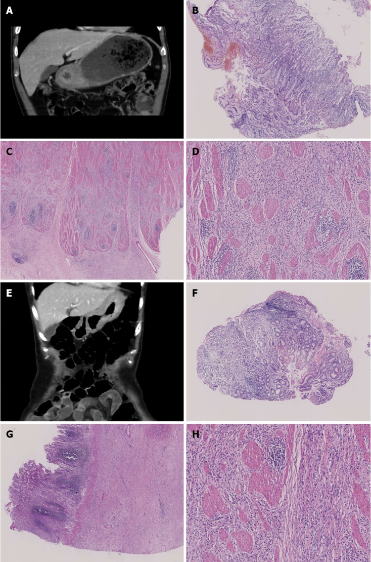
Figure 1.

Computerized tomography and pathological findings. A: Diffuse thickened antral detected by computerized tomography (CT) scan; B: Repeated false negative slides without cancer cells detected for the previous 5 biopsies [hematoxylin-eosin (HE) stain, × 40]; C: Positive slide with cancer cells observed by the 6th biopsy (HE stain, × 100); D: Postoperative slide with cancer cells observed, many signet ring-like cells can be observed in the upper-right quadrant of the slide (HE stain, × 200); E: Diffuse thickened fundic and gastric body detected by CT scan; F: Repeated false negative slides without cancer cells detected for 8 biopsies (HE stain, × 40); G: Postoperative slide with cancer cells observed and the whole gastric wall infiltrated (HE stain, × 100); H: Fibrosis stomach wall, scattered or small focal distributed signet ring-like cells could be observed (HE stain, × 200).
