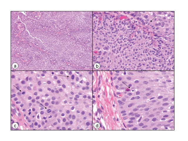
Fig. 8.

Microphotograph of the papillary urothelial neoplasm of low malignant potential showing discrete and non-fused papillae, which are lined by multilayered urothelium with minimal to absent cytologic atypia. There is an increase of cell density as compared to normal urothelium (A). However, the polarity, particularly of the basal layer (B, arrow), is preserved with palisading and an impression of predominant order is given. Obviously, there is also minimal variation in architectural and nuclear features and the nuclei are enlarged as compared to normal urothelial cells (B). Mitoses are rare, but are occasionally seen, although are not atypical (C, arrows). An apoptotic body is also seen (D, arrow) (Hematoxylin-eosin staining, a: ×100, b: ×400, c: ×630, d: ×630).
