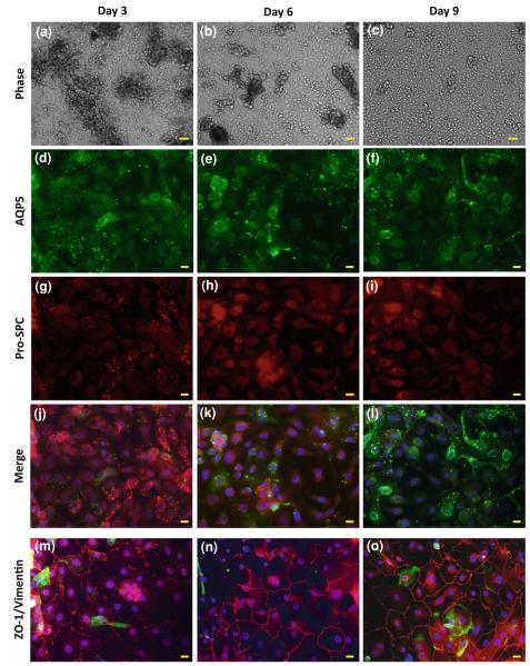
FIGURE 2.
Freshly isolated human alveolar epithelial cells were cultured on collagen coated Silastic membranes, and phase images were taken after (a) 3, (b) 6, and (c) 9 days in culture. On the same days, the cells were fixed and immunostained for (d–f) AQP5, a type I AEC marker, and (g–i) pro-SPC, a type II AEC marker. (j–l) Merge of the AQP5/pro-SPC stains with DAPI stain for the nuclei (in blue). Other cultures that were fixed after (m) 3, (n) 6, and (o) 9 days in culture were stained for ZO-1 (red), vimentin (green), and the nuclei were stained with DAPI (blue). Bar for phase images = 50 μm; Bar for immunofluorescent images = 10 μm.

