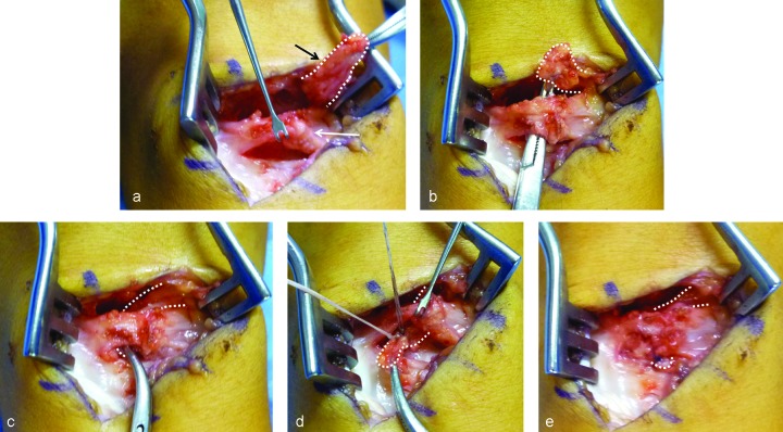
Fig. 2a–e.
Surgical technique of dorsal capsulodesis by using a short skin incision with extensor retinaculun preservation. (a) Capsule exposure with maximum preservation of the capsule itself (white arrow) using a double almost-parallel capsular incision. The proximal incision is on the dorsal radius, and the second one is on the midcarpal joint following the direction of the dorsal intercarpal ligament (DICL) fibers. After synoviectomy, a DICL flap fixed at the scaphoid is elevated (white dotted line). (c) A tunnel space is created under the capsule over the SL ligament level and a curved mosquito is passed to keep the DICL flap. (c) The DICL flap is passed under the capsule through the tunnel and (d) is fixed to the dorsal border of the lunate with a suture anchor. (e) Capsule is sutured at the end.

