Fig. 4.
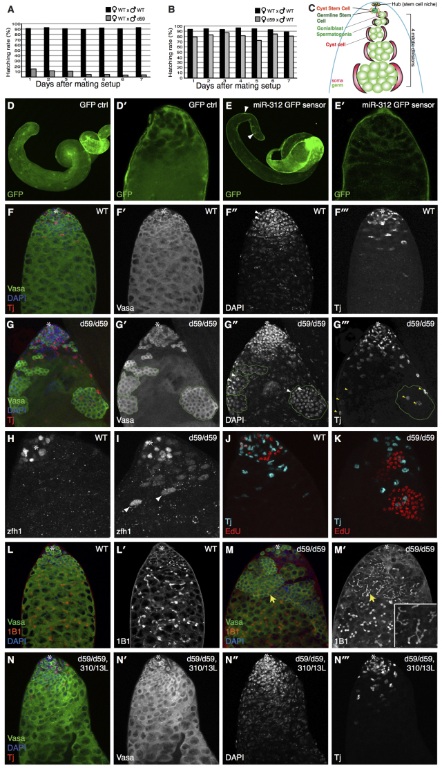
miR-310/13 null flies (d59/d59) exhibit a male-specific fertility defect. (A,B) d59/d59 males exhibit severe sterility. Hatching rate of eggs laid by d59/d59 males (n=200) crossed to WT females is significantly decreased (gray bars) compared with a WT control cross (black bars) (A). d59/d59 females exhibit no significant fertility defect (B). (C) Model of WT testes (see text for details). (E,E′) GFP sensor containing multimerized miR-312 binding sites displays a marked reduction of the GFP signal in the apical and medial domain of the testis, but not in the nuclei of the muscle sheath cells (arrowheads in E; see quantification of GFP sensor fluorescence intensity in supplementary material Fig. S2I-K). (D,D′) Note that there is increased cytoplasmic GFP expression in both soma and germ in the control testis. However, we consistently see much weaker GFP expression in germ cells compared with soma. (F-F′′) WT testis showing the germline marker Vasa in green, DAPI in blue, and the CySCs/early somatic cyst cell marker Tj in red. (G-G′′) Testes of d59/d59 flies exhibit abnormal accumulations of early germ cells and somatic cyst cells. The germ cell clusters (outlined by green dashed line in G′-G′′) accumulate further away from the hub compared with WT control. The cells within these clusters display condensed nuclear morphology, as shown by DAPI staining (arrowheads in G′) and appear to be poorly differentiated. The germ cell clusters are almost always associated with ectopic Tj+ cells (G,G′′), which show a marked increase in number compared with WT (compare G′′ with F′′). (H,I) Ectopic cyst somatic cells are positive for the CySC/early cyst cell marker Zfh1 in d59 mutant testes (I), compared with WT (H). Note that the bright Zfh1+ cells (arrowheads in I) are associated with germ cell clusters (shown in the merge in supplementary material Fig. S2M). (J,K) Synchronous cell division in abnormal germ cell clusters in d59/d59 testes (K), compared with WT (J), as revealed by EdU staining. (L-M′) Abnormal germ cell accumulations in d59 homozygous testes harbor branched fusomes (M′). 1B1 marks fusomes (red) (L,M). GSCs contain ‘dot’ fusomes, and the degree of fusome branching in germ cells correlates with the extent of differentiation (L,L′). miR-310/13-deleted testes display extensive branched fusomes within abnormal germ cell clusters (yellow arrows in M,M′; higher magnification in M′ inset). (N-N′′) Rescue of the germ cell accumulation phenotype in d59/d59 males expressing a genomic construct spanning the mir-310-313 locus as well as a region upstream of the transcription start site (miR-310/13L). Asterisks mark the hub.

