
Fig. 2.
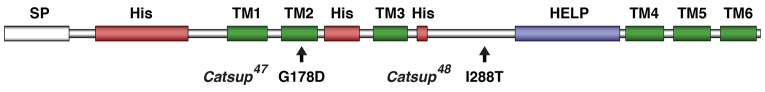
Structure of Drosophila Catsup protein and locations of the Catsup47 and Catsup48 mutations. Catsup protein showing the predicted signal peptide (SP), histidine-rich regions (His), conserved HELP domain (Suzuki and Endo, 2002) and six transmembrane domains (TM1-6). Locations of amino acid alterations in the newly isolated mutants Catsup47 and Catsup48 are indicated.