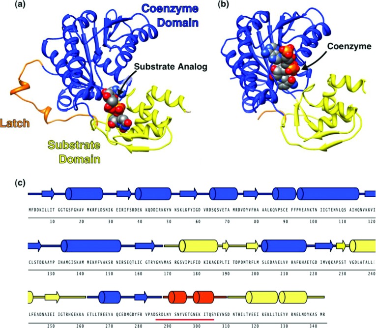
Figure 2. Crystal structure of CapE.
Structure of (a) CapE with substrate analogue UDP-6N3-GlcNAc bound, and (b) K126E mutein with coenzyme NADPH bound. The coenzyme-binding domain, substrate-binding domain, and latch are coloured in blue, yellow and orange, respectively. The ligands are depicted as spheres with CPK colours. The figure was prepared with CHIMERA [50]. (c) Primary and secondary structure of CapE. The underlined sequence (red line) corresponds to the disordered region in K126E.

