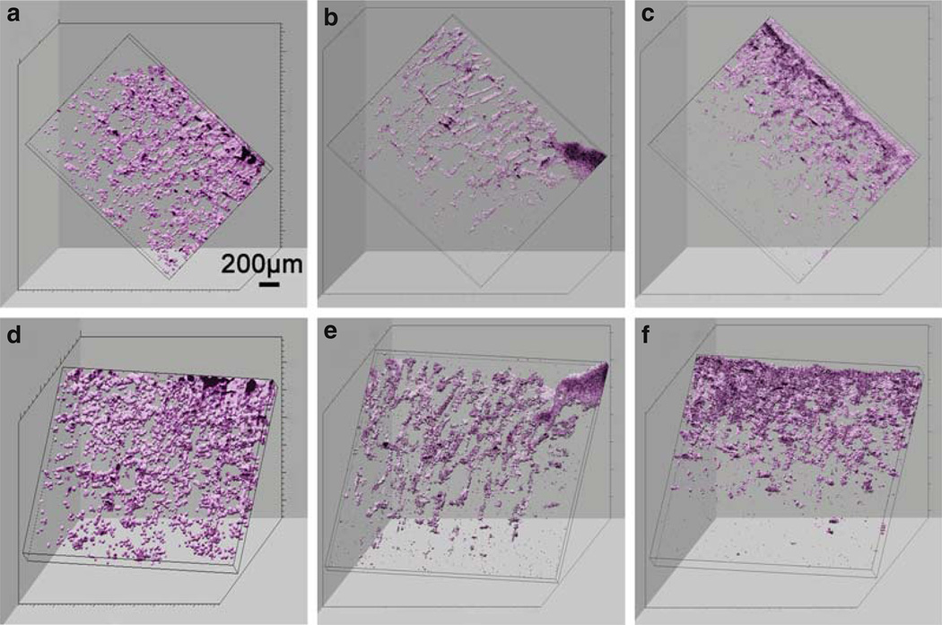
Fig. 13.3.
Three-dimensional OCT reconstructions of chitosin scaffolds from two different viewing angles taken at 3 (a, d), 5 (b, e), and 7 (c, f) days. Rotational angles of image a–c are 40° (y), 20° (z), 70° (x) while d–f are 0° (y), 10° (z), 50° (x). Viewing a 3D reconstruction from many angles, and computationally sectioning out planes from arbitrary angles, enables a more complete view and assessment of the 3D characteristics of a sample, compared to single cross-sectional images. Modified figure used with permission (20).

