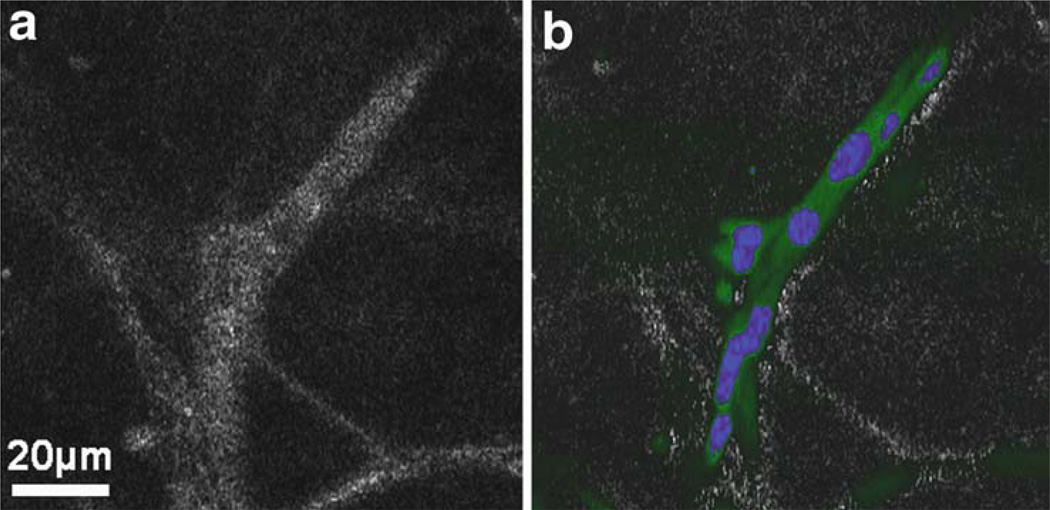
Fig. 13.6.
OCM (a) and MPM overlaid on OCM (b) images from a 3D Matrigel scaffold seeded with GFP-vinculin fibroblast cells stained with a nuclear dye prior to mechanical stimulation. The green signal in the MPM image is from GFP and indicates the relative degree of cell adhesion. The blue signal is the nuclear stain which identifies individual cells. Scattering in this culture, as seen in the OCM image, is caused by both the cells and the extra-cellular environment (collagen secreted by the cells). It is observed that the cells have organized into an interconnected architecture along the fibrous collagen structure of the Matrigel. Modified figure used with permission (31).

