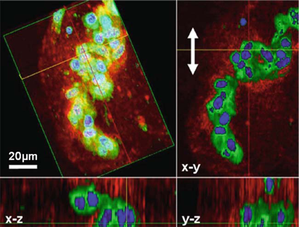
Fig. 13.7.
Multimodality 3D OCM/MPM image of GFP-vinculin fibroblasts in a 3D Matrigel scaffold acquired after mechanical stimulation. The 3D reconstruction is shown (top left) as well as projections along different axes giving a comprehensive view of this cluster of cells. The red channel corresponds to the scattering from the sample (OCM) while the green and blue channels are fluorescence images (MPM). Three-dimensional reconstruction, rotation, and computational sectioning at arbitrary plane angles allow for a more comprehensive view of a 3D cell culture. Mechanical stimulation in the direction of the arrows in the x-y plane has induced the cells to become more spherical and form clusters, potentially indicating that adhesion sites with the scaffold were broken during mechanical stimulation. Modified figure used with permission (31).

