
Figure 2.
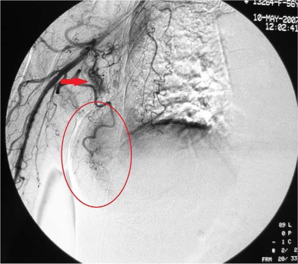
Digital subtraction angiography of the right subclavian artery.
Notes: Digital subtraction angiography reveals that the lateral thoracic artery, which supplies the tumor, becomes thickened and tortuous. The tumor appears patchy, with an approximate diameter of 7 cm, as indicated by the red oval. Multiple lymph nodes appear nodular and are supplied by numerous smaller branches of the subclavian artery.